New MRI-Guided Radiation Therapy System Puts Us on Target to Deliver Therapy With Greater Precision
If you’ve seen Top Gun or the new Maverick movie, then you’re probably familiar with the term “missile lock.” It’s that sweet spot, when your target is in your sights and perfectly aligned for a successful hit. For our radiation oncologists, their target, of course, is cancer. And now, they have a new tool that will make it easier than ever to hit their mark: the MR-linac.
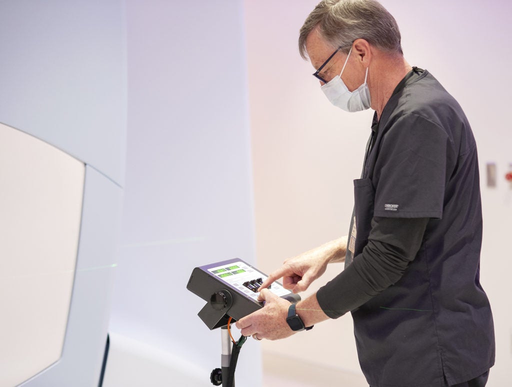
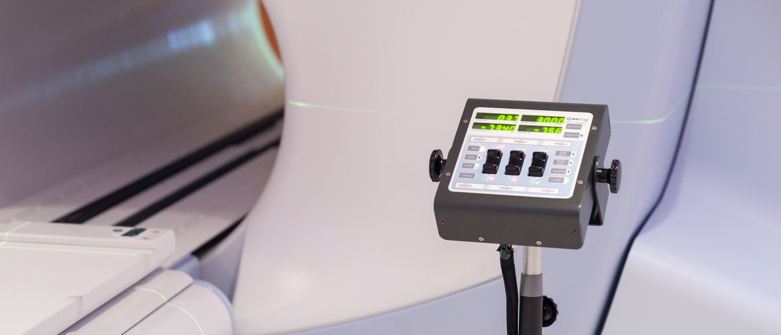
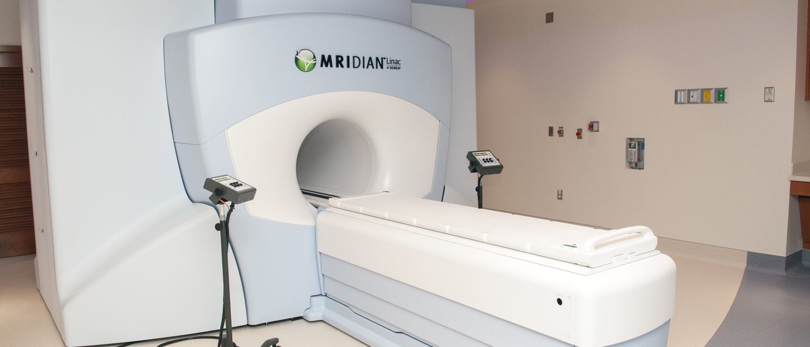
The MRI-guided radiation therapy system, MRIdian, combines the superior visualization of magnetic resonance imaging (MRI) and a linear accelerator into one innovative device that can deliver high-dose radiation with greater precision. “The combination of these two technologies really allows us to do amazing things that we couldn't do before,” says radiation oncologist Einsley Janowski, MD, PhD. “It’s really a huge step forward in maximizing both patient outcomes and patient quality of life.”
Why the MR-Linac Is a Gamechanger for Cancer Care
Radiation therapy has been used for over a century to treat cancer, and the technology has continued to evolve to boost cure rates and limit side effects. The MR-linac is the most recent evolution designed to help overcome some of the limitations of conventional radiation therapy. It offers:
- Superior imaging | An X-ray or CT scan is traditionally used to help pinpoint the location of a tumor prior to radiation therapy. An MRI provides a more detailed image and can help doctors distinguish between soft tissue and a tumor. According to Paul Read, MD, medical director of the Department of Radiation Oncology, the more clearly we can see the margins of the tumor, the better our chances of avoiding normal tissue during treatment. The result: fewer side effects.
- Better aim for moving targets | Tumors can change shape or move during treatment, particularly in areas of the body that shift with respiration (e.g. the lungs) or with digestion (e.g. the abdomen). The MR-linac allows the radiation oncologist and radiation therapist involved in each treatment to see the tumor in real time during the therapy. This means they can lock in and radiate the tumor when it falls into the target area, and they can turn off the radiation when the tumor is not aligned properly.
“In the past, radiation therapy was like aiming our gun at the target, then closing our eyes and pulling the trigger. Now, we're going to be able to keep our eyes open as we pull the trigger and keep them open throughout the radiation treatment,” says Director of Radiological Physics Jeff Siebers, PhD.
- Treatment flexibility | Real-time visualization also means that the care team can adapt the treatment to account for real-time changes in the body. “We can do a daily adaptive plan to personalize the cancer treatment for that patient's particular anatomy on that particular day,” says Janowski. “What used to take us a week to plan, we can literally do while the patient is on the table. We can adapt the treatment so that we can precisely focus on the tumor and maximize the radiation dose, but avoid the adjacent structures that could cause toxicity in the future.”
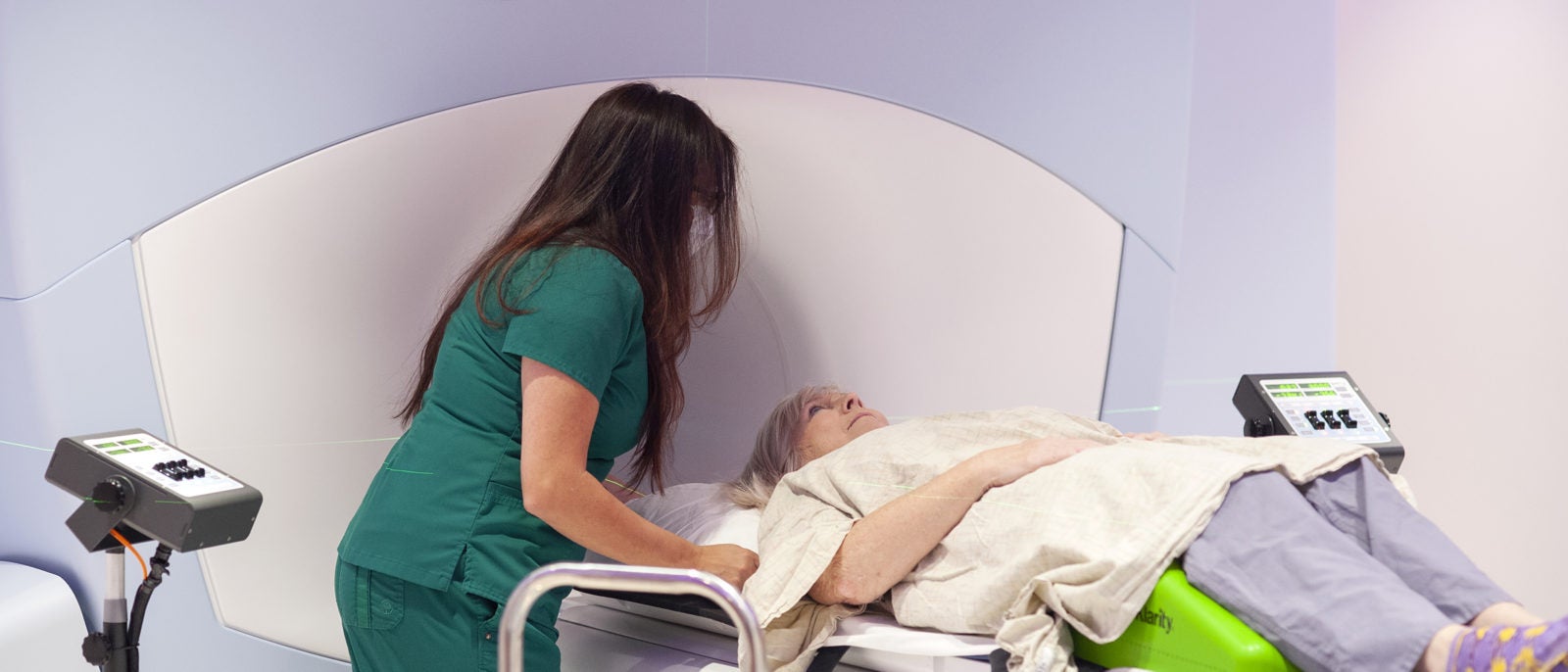
- Patient involvement | The treatment experience for the patient will essentially be the same as traditional radiation therapy, according to Joan Harris, RN. However, there is one key difference: the MRIdian has a video screen that allows the patient to see what’s happening so that they can participate in their treatment. “The patient can see the tumor position and can adapt their respiration to be sure it stays in the target area during treatment,” says Janowski.
“A lot of people compare it to a video game,” adds Laura Bauchert, customer success manager with device vendor, ViewRay. “You can see your anatomy and where the tumor is. They get a smiley face when they get the tumor in the target. They get positive feedback, and they’re able to see where exactly they’re being treated.”
Which Patients May Benefit Most from MR-Linac Treatment?
Like any medical device, the goal is to use the MR-linac for those patients we know this technology will benefit most. “Almost any tumor can be treated with an MR-linac,” says Read. “But it is best used for patients who have tumors in a position where there is movement like the abdomen.”
“There are a variety of different judgements that will go into determining who’s going to be best treated with this technology,” adds Janowski. “It’s going to be another tool in our arsenal and it will allow us to be aggressive in treating some of those cancers like pancreatic cancer that are notoriously difficult to treat.”
Patients with lung, liver and prostate cancer also will be potential candidates for MR-linac treatment. Yet, this list is growing as more centers use this system. “In time, we anticipate that patients with many other cancers will benefit from MR-linac treatment,” says Read. And UVA Cancer Center will be on the forefront of efforts to advance the use of this technology further through participation in clinical trials. The first up will be trials for patients with rectal cancer, followed by pancreas and prostate cancers.
Plus, Read says: “Our research teams will have access to daily MR scans during cancer treatment, which has never been possible before. They can study these images to see if they can learn how cancers respond to treatment, and if we can use the information to predict who will be cured and to determine why some patients have more radiation side effects.”
How Many Team Members Does It Take to Facilitate MR-Linac Treatment?
The short answer: a lot. But radiation oncology has always been a team sport, says Janowski. Incorporating this new treatment into their playbook has taken some planning and practice and some additional training, but all key players are now ready to do their part. They include:
- Radiation oncologists who oversee every aspect of the patient’s care, including determining which treatment is the best option, and planning how much radiation is needed and where it should be delivered.
- Physicists and dosimetrists who plan the treatment and ensure the right dose of radiation is delivered to the tumor while avoiding healthy tissue. They are also responsible for quality control, and optimizing the technology to maximize its benefits.
- Nurses who educate the patients on the treatment and necessary safety measures, help minimize side effects of treatment and provide care and support throughout the treatment process.
- Radiation therapists who perform the MRI scan, position the patient appropriately, administer radiation treatment and counsel the patient throughout to help ensure the best outcome.
What will be unique about the MR-linac treatment process compared to our traditional radiation treatment is the way these key players collaborate on the day of treatment. The ability to adapt the treatment plan in real time due to shifts in the tumor, organs, or other bodily changes means that everyone has to be at the ready when the patient goes into treatment. “We're going to be working very closely together instead of handing the various tasks off person to person like a baton. We're all going to be running this race together to get the treatment done as quickly as we can while the patient is on the table,” says Janowski.
Why Is UVA Cancer Center a Perfect Home for This Technology?
“Very few centers across the world have this technology,” says Read. In fact, only around 25 have been installed nationwide, and UVA Cancer Center is one of only two centers in Virginia to make this part of their cancer-treating arsenal.
We are the only NCI-designated, comprehensive cancer center in the state. We have a proven commitment to advancing cancer treatment, the solid foundation necessary to bring this kind of innovative technology to our patients, and the ability to provide patients the comprehensive care they need at every turn, from diagnosis to recovery.
“We have this really great infrastructure of dedicated and brilliant oncologists, and compassionate and capable nurses and support staff. We provide multidisciplinary care with a terrific collaborative approach focused solely on providing the best outcomes for our cancer patients,” says Janowski. “This is another new, exciting tool that's just going to allow us to offer even more options for our patients and I think, hopefully, push the field forward.”
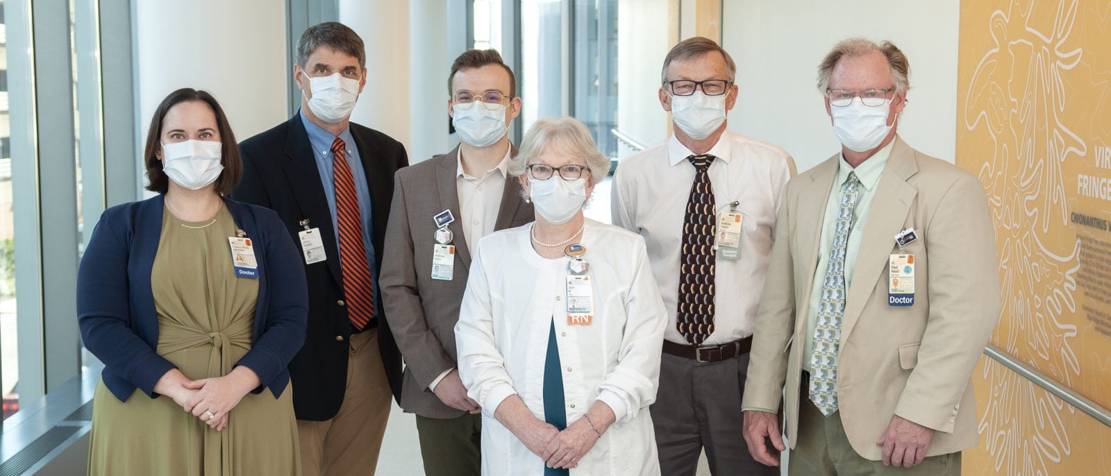
(l-r) Dr. Einsley-Marie Janowski, Dr. Jeff Siebers, Matthew Mistro, Joan Harris, Andrew Taylor, and Dr. Paul Read.
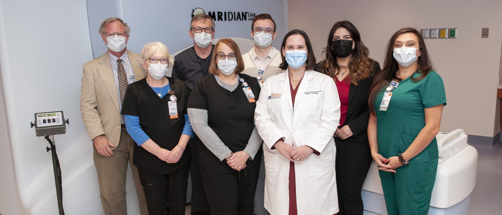
Front row (l-r): Joan Harris, Johanna Ellwood, and Dr. Einsley-Marie Janowski.
Back row (l-r): Dr. Paul Read, Andrew Taylor, Matthew Mistro, Tatiana Bejarano, and Mariana Miller.
*****
What does it take to install a 20-foot-long, 19-foot-wide, high-tech device into a busy cancer center during a pandemic and a supply shortage? Take a look at how we did it!
Latest News

